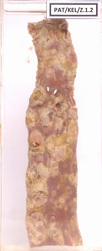
Z.1.2 Atheroma of artery
- The whole luminal surface of aorta appears grossly distorted.
- There are irregular raised yellowish patches which are the atheromatous plaques.
- In between the plaques certain areas of the intima appear thickened and raised above the surface.
- Some atheromatous plaques have ulcerated.
Clinical presentation
The specimen displays the descending aorta of a 70-year-old man who died following a congestive cardiac failure
Questions
- Describe the lesions that are observed in the aorta.
- Describe the evolution and complications of these lesions.
- Name three complications this man would have developed due to these lesions found in his descending aorta.
Answers
1. Describe the lesions that are observed in the aorta.
- Complicated atheromatous lesions including irregularly shaped plaques with surface ulcerations.
- Located around the ostia of the branches of the descending aorta.
- Possible thrombosis is noted in the upper lesions.
2. Describe the evolution and complications of these lesions
- Start as fatty streaks in adolescence/ young age by deposition of lipid laden foamy macrophages coalescing to form streaks. These are not sufficiently raised up to cause a disturbance in vascular flow.
- Develops into atheromatous plaques due to lipid deposition and intimal thickening forming an elevated lesion. These coalesce to form enlarging plaques that cause disturbance of the flow.
- Plaques generally continue to change and progressively enlarge. These are subjected to various changes and remodelling.
Eg: calcification, fibrous thickening, organization of superimposed thrombi
- Complications:
- Rupture, ulceration, or erosion of the surface of atheromatous plaques exposes highly thrombogenic substances and leads to thrombosis, which may partially or completely occlude the vessel lumen
- Haemorrhage into a plaque- Rupture of the overlying fibrous cap, or of the thin-walled vessels in the areas of neovascularization, can cause intraplaque haemorrhage; a contained hematoma may expand the plaque or induce plaque rupture.
- Atheroembolism- Plaque rupture can discharge atherosclerotic debris into the bloodstream, producing microemboli.
- Aneurysm formation. Atherosclerosis-induced pressure or ischemic atrophy of the underlying media, with loss of elastic tissue, causes weakness and potential rupture.
3. Name three complications this man would have developed due to these lesions found in his descending aorta.
- Distal thromboembolism - peripheral vascular disease, mesenteric infarctions
- Abdominal aortic aneurysms and rupture of aneurysms
- Renal artery stenosis -due obstruction of the orifice
Microscopy
Briefly describe the microscopy of an atheromatous plaque
- Superficial fibrous cap composed of smooth muscle cells and relatively dense collagen.
- Beneath the cap, is a more cellular area containing macrophages, T cells, and smooth muscle cells.
- Deep to the fibrous cap is a necrotic core, containing lipid (primarily cholesterol and cholesterol esters), debris from dead cells, foam cells (lipid-laden macrophages and smooth muscle cells), fibrin, variably organized thrombus, and other plasma proteins.
- The periphery of the lesion demonstrates neovascularization
Links to images
Microscopic appearance of an atheromatous plaque 1 - high power
Microscopic appearance of an atheromatous plaque 2 - high power
Microscopic appearance of an atheromatous plaque - low power