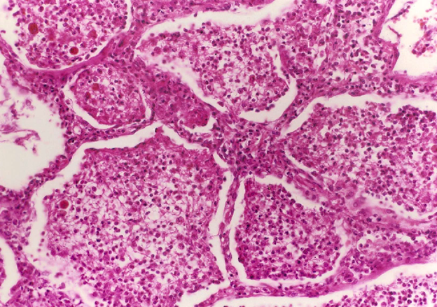
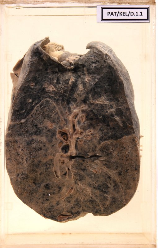
D.1.1 Lobar pneumonia
- The cut surface shows the left lung with the hilum containing pulmonary blood vessel and bronchi.
- Note the clear demarcation between upper and lower parts of the lung.
- Upper dark area is normal lung tissue.
- Lower part - grey hepatisation.
- Pleural surface shows fibrin tags.
Clinical description
A 50-year-old male with diabetes mellitus had an episode of fever and productive cough and died on the fifth day of the illness. The specimen shows his left lung.
Questions
1. What is the reason for the grey colour in the lower lobe?
2. What is the reason for the formation of fibrin tags on the pleural surface?
Answers
1. What is the reason for the grey colour in the lower lobe?
The grey colour is due to dense fibrin strands
2. What is the reason for the formation of fibrin tags on the pleural surface?
There will be pleuritis
↓
Fibrinopurulant exudate accumulation within the two sheaths of pleura
↓
This exudate gets organized
↓
There will be adhesions between the two sheaths of pleura
↓
Following the separation of the two sheaths broken adhesions will appear as fibrin tags