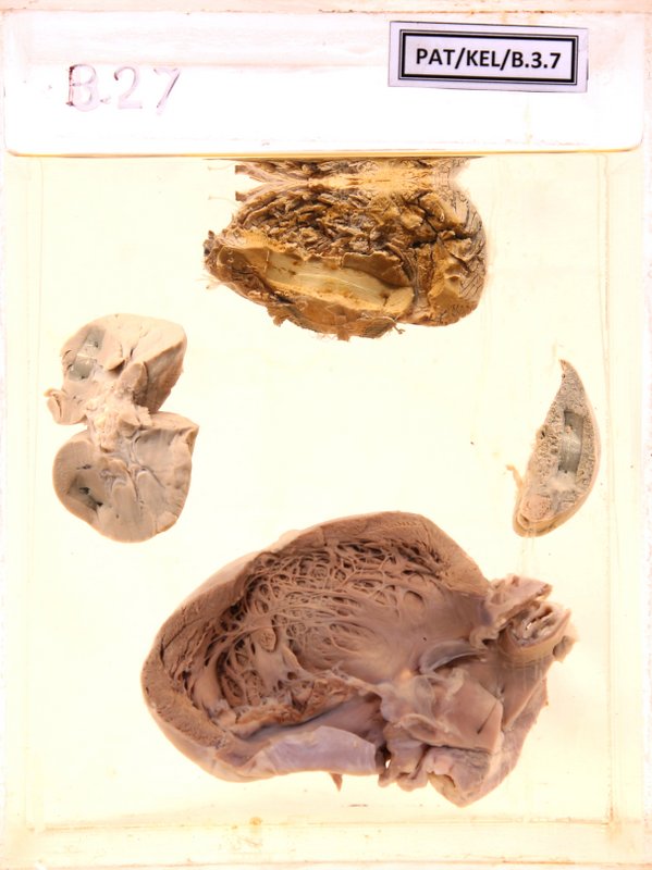
B.3.7 Infective endocarditis with infarcts in multiple organs
- The left ventricle, left atrium and the aorta are opened into.
- On the mitral valve and on the papillary muscles there are warty vegetations.
- The mitral valve cusps are destroyed.
- The kidney and spleen show infarcts and the brain shows some necrosis.
Clinical description
The specimen displays parts of organs resected at the postmortem of a 35-year-old woman who died while investigating for pyrexia of unknown origin (PUO).
Questions
1. Describe the macroscopic changes observed in the mitral valve cusps and related structures.
2. Describe the microscopy of the valvular lesion
3. What are the possible causes of death?
4. What is the most likely underlying cardiac disease that has contributed to her current illness?
5. Briefly describe the pathogenetic basis of the lesions that you observe in the other organs ?
Answers
1. Describe the macroscopic changes observed in the mitral valve cusps and related structures.
The mitral valve cusps are distorted with irregular and nodular surface
Nodular and irregular deposits on the atrial surface of the mitral valve- Vegetations
The chordae tendinae of the mitral valve appears thickened and irregular
The papillary muscles appear thickened
2. Describe the microscopy of the valvular lesion
Please see tab on "Microscopic appearance"
3. What are the possible causes of death?
Septicaemia with multiorgan failure/septic shock
Cardiac failure
Cardiorespiratory failure due to brain herniation
4. What is the most likely underlying cardiac disease that has contributed to her current illness?
Rheumatic valvular disease
5. Briefly describe the pathogenetic basis of the lesions that you observe in the other organs ?
The friable vegetations dislodged due to rapid stream of the blood and give rise to embolization in the systemic circulation and affect organs like spleen, kidney and brain causing infarcts and abscesses
Microscopic apearance
The microscopy of the valvular lesion
- The outer most layer consists of eosinophilic material composed of fibrin and platelets.
- Beneath that there are colonies of microorganisms that appear basophilic.
- The underneath valvular stroma is oedematous with an infiltrate of chronic inflammatory cells, scattered neutrophils and inflammatory granulation tissue formation.