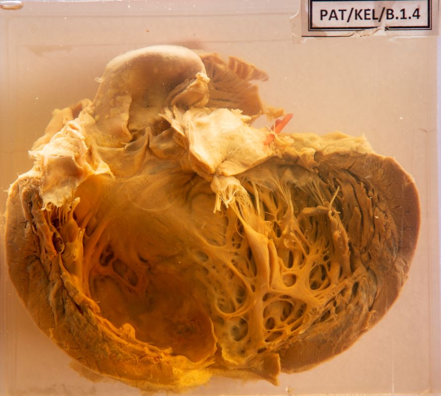
B.1.4 Old myocardial infarct with aneurysmal dilatation of the apex
- The aorta shows atheromatous plaques just above the aortic cusps.
- The left ventricular wall shows hypertrophy.
- At the apex, on the pericardial and the endocardial surfaces there are blotchy areas.
- The white blotches probably are either fibrosed or calcified spots.
- There is aneurysmal dilatation of the wall at the apex.
- The coronary arteries show narrowing and are of irregular caliber.
Clinical presentation
A 68-year-old man was admitted to the emergency unit with severe chest pain for one hour. Despite treatment, he died eight hours following admission. He is a known patient with poorly controlled hypertension for 15 years. He has a history of a myocardial infarction one year ago.
A postmortem was performed.
The specimen (B 1.4) displays the cut opened left ventricle of his heart.
Macroscopic changes
Describe the macroscopic changes you observe in his heart.
- There is left ventricular wall hypertrophy.
- The apex shows aneurysmal dilatation (see Arrow, Image 1) and pale colour myocardium, probably fibrosed foci related to the previous infarct.
- Atheromatous plaques are noted just above the aortic cusps.

Image 1 - Aneurysmal dilatation