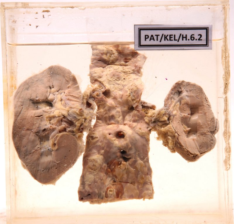
H.6.2 Granular contracted kidneys due to atheromatous changes involving the renal artery
- Specimen shows both kidneys with abdominal aorta.
- Right kidney surface appears smooth.
Cut surface
- Cortico – medullary demarcation is not distinct.
- Cortex shows few white streaks probably due to scarring.
Left kidney
- Shows marked atrophy.
- The surface is granular.
- The cortex is thin and the medulla shows white streaking probably due to fibrosis.
- The left renal artery shows stenosis and almost complete obstruction due to atheromatous changes.
- Probably the atrophy of the left kidney is due to the renal artery stenosis.
Aorta
- The right renal artery has a thick wall. The inner surface of the aorta shows yellow colour irregular atheromatous plaques.
- Some of the plaques show ulceration.