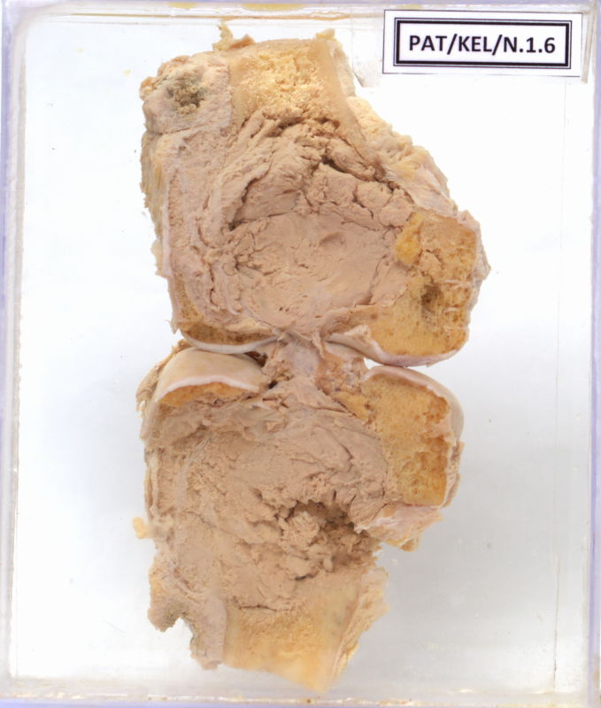
N.1.6 Osteoclastoma – Lower end of femur
- From a 52 year old male patient who presented with a history of lump in the left side femur.
- The outer surface shows the condyles of the lower end of the femur covered partially by fibrofatty tissue.
- The cut section shows markedly thinned out bony and cartilaginous margins enclosing a pale pink rather homogeneous soft tumour tissue.
- The tumour appears to have invaded the marrow space.
- The right of the tumour shows necrotic tissue.
- The bony tissue beneath that shows cystic changes probably due to osteolystic changes and appears pale yellow in colour.