K.6.1 Vulval tumour
- The specimen of a lump at the vulva of a 21 year old female patient who presented with a history of a painful swelling at the vulva of 3 months duration.
- Clinical examination showed a large fleshy growth of the left labium majus, with haemorrhage into it.
- At surgery the growth was shelled out of its covering.
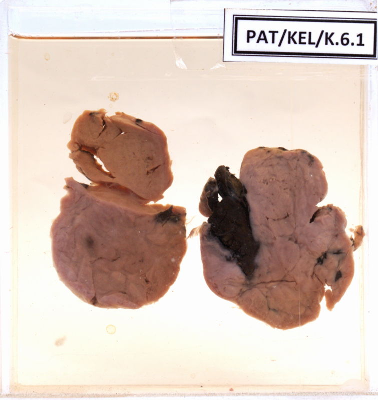
- The specimen shows an oval shaped firm lesion.
- The surface is lobulated, with patchy, dark (haemorrhagic) areas.
- The cut section has a solid, white appearance, and is lobulated.
- Lobules are of varying sizes.
- Some of the small lobules have a whorled appearance.
- A large area of organized blood and many haemorrhages were seen on cut section.
- Differential diagnosis include
Hidradenoma
Chronic bartholin cyst
Fibroma of the vulva
- Histology confirmed a Leiomyoma of the vulva.