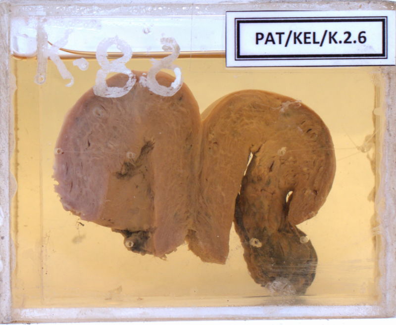
K.2.6 Placental Polyp
- The specimen is from a 38 years old patient who presented with bleeding per vaginum for 7 weeks duration after a LSCS.
- An abdominal hysterectomy was done.
- The specimen is that of the uterus cut coronally and shows a large polypoidal mass arising from the right cornu of the uterine cavity projecting down to the cervix.
- Haemorrhagic and necrotic areas can be seen in the lower part of the growth.
- Histology showed placental tissue with calcification.