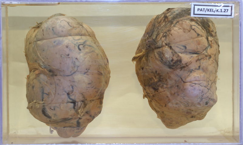
K.1.27 Brenner tumour of the ovary
- 62 years old female admitted with a lump in the abdomen.
- She has a past Hx of total abdominal hysterectomy and left salphingo oophorectomy done 27 years ago for leiomyoma of the uterus.
- At laparotomy a solid lump measuring 15×11×8 cm removed.
- Adhesions to omentum present. No ascites.
- The right ovary shown as irregular lobulated outer surface.
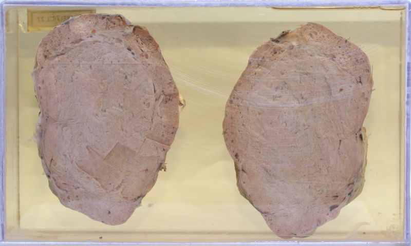
- Cut inner surface shows a fairly well demarcated area which appears solid with the periphery of the tumour showing cystic areas.
- A large cyst filled with brown coloured material seen.
- Histology showed a solid tumour composed of nests of epithelial cells in a dense fibroblastic stroma and microcyst formation, confirming a Brenner tumour of the ovary.