This is descending aorta of a 57 year old male, presenting with abdominal distension for 2 days.
On examination he was pale with ascites.
At the exploratory laparotomy there was retroperitoneal haematoma on the left side.
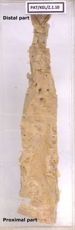
In the specimen the aorta has been cut open to show the descending part, up to its bifurcation.
Along the whole length, it shows, yellow colour material beneath the intima – atheromatous plaques with foci of marked calcification, ulceration and superadded thrombosis.
The media is thinned out, especially at the bifurcation of the aorta.
Above the bifurcation there is a stenosed part identified at the post – mortem (indicated by an arrow).
Here the adventitia shows brownish staining - ?Tracked down blood, from severely ulcerated, atheromatous plaque