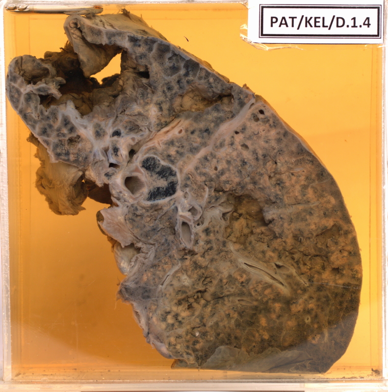
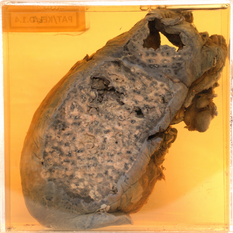
D.1.4 Tuberculosis
- The pleural surface of the lung shows patchy, white areas.
- The upper part contains a large cavity in the apex.
- The floor of the cavity is lined by fibrous bands, and is covered by dark necrotic material.
- However the walls appear fairly smooth (Chronic cavity).
- Smaller cavities can be seen throughout the lung, with ragged, caseating walls.
- One of these (top left) appears to have opened into pleural cavity.
- The specimen also has small broncho pneumonic patches, especially in the lower part.
Diagnosis - Progressive secondary tuberculosis with an apical cavity and acute tuberculous bronchopneumonia.