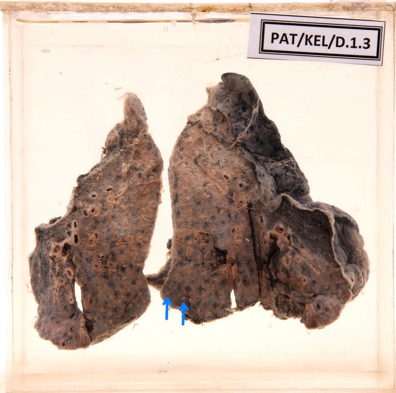
D.1.3 Tuberculous bronchopneumonia
- The cut surface shows a grey colour area of consolidation with dark carbon mottling.
- Minute whitish - tan coloured mottling (probably tubercles) scattered throughout lung tissue.
- A small portion of the lung in the periphery appears normal.
- The pleural surface shows fibrinous tags and whitish consolidation through the pleura.