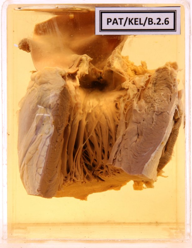
B.2.6 Left ventricular hypertrophy due to aortic stenosis
- The left ventricle is cut opened.
- Marked hypertrophy of the left ventricle and the papillary muscles.
- The aortic cusps show fusion and stenosis.
- The aorta shows few atheromatous patches and post stenosis dilatation.
- The right ventricle is just slit open.